

Ultrasound Training Simulator for Medical Students: Revolutionizing Clinical Education
In the rapidly evolving landscape of modern healthcare, point-of-care ultrasound (POCUS) has emerged as an indispensable diagnostic tool. Often referred to as the stethoscope of the twenty-first century, it provides immediate, non-invasive imaging directly at the patient's bedside. However, acquiring the psychomotor skills and cognitive spatial awareness necessary to interpret these dynamic images is notoriously difficult. To bridge this profound educational gap, academic institutions globally are rapidly integrating the ultrasound training simulator for medical students into their core curricula.
Historically, learning sonography relied heavily on an apprenticeship paradigm, where medical students practiced on live patients or healthy volunteers. Today, relying strictly on live human practice is no longer ethically or logistically viable for foundational learning. By heavily utilizing an ultrasound simulator, universities ensure that future physicians can develop a robust baseline of clinical competence in a completely safe, highly controlled environment. The deployment of a dedicated ultrasound training simulator for medical students guarantees that learners can make mistakes, refine their probe positioning, and profoundly improve their image interpretation skills before they ever touch a critically ill patient.
The Paradigm Shift: Ultrasound Simulation Models Training
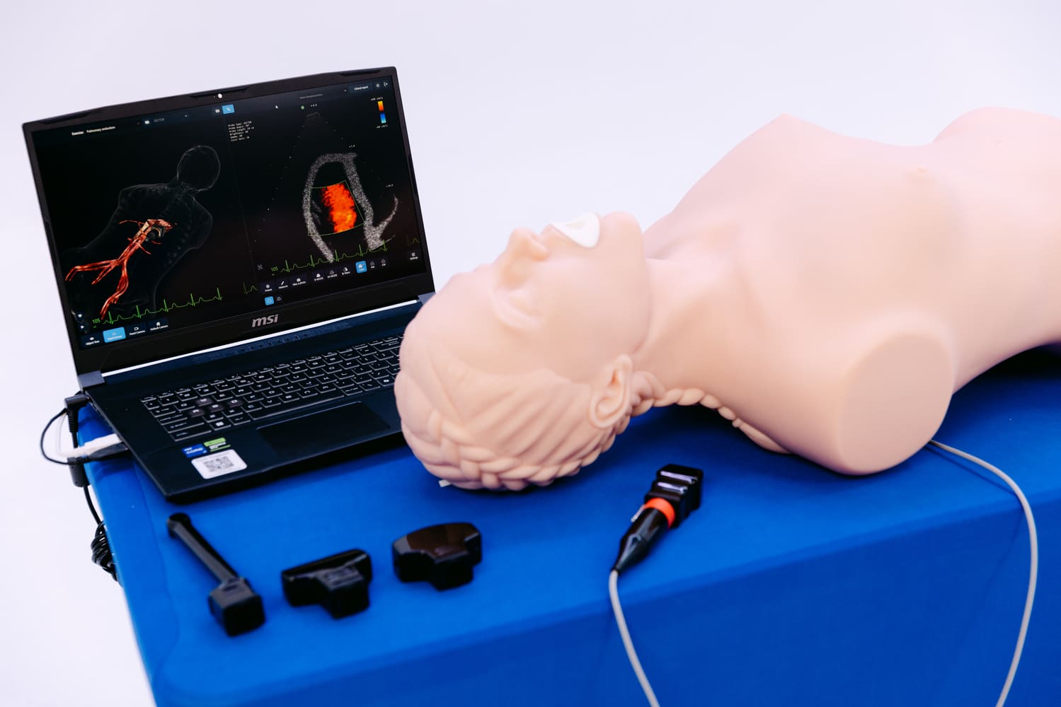
The transition from traditional textbook education to immersive, technology-based experiential learning requires sophisticated engineering. Advanced ultrasound simulation models training perfectly embodies this paradigm shift. An effective ultrasound simulation model is not merely a plastic mannequin; it is a highly complex, technologically integrated system that flawlessly replicates the physical and acoustic properties of the human body.
When academic directors evaluate ultrasound simulation models training, they generally categorize these systems into two distinct types of simulators: physical phantom models and high-fidelity virtual reality (VR) simulators.
Physical Phantom Models
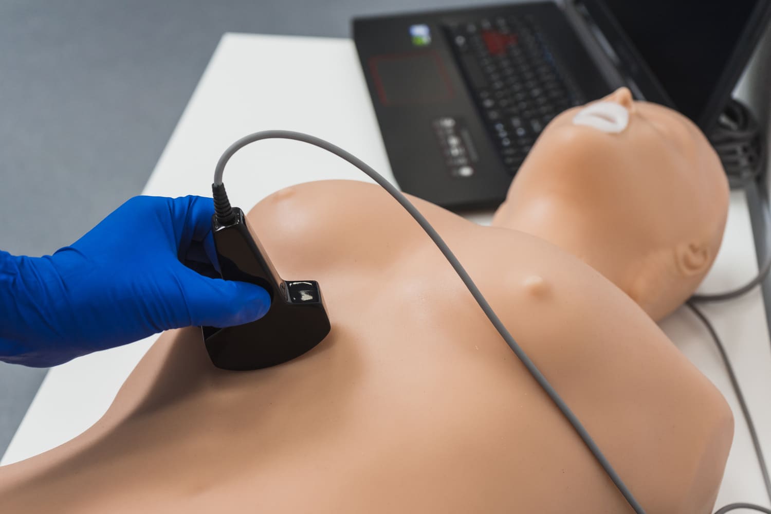
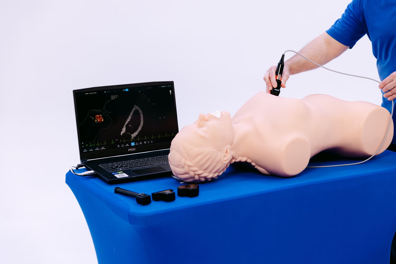
A physical phantom model is constructed from specialized, tissue-equivalent materials that interact with actual high-frequency sound waves exactly like human flesh, fat, and bone. During ultrasound simulation models training, medical students use a real, functioning ultrasound machine and a real transducer probe. When they apply acoustic gel and press the probe against the synthetic model, the screen displays a genuine ultrasound image generated in real-time. This specific type of simulator is absolutely vital for teaching tactile feedback. Students learn exactly how much physical pressure to apply to the model to obtain a clear image without compressing delicate underlying veins.
Virtual Reality and Augmented Reality Simulators
Conversely, virtual reality simulators utilize an artificial, computerized probe paired with an interactive digital screen or VR headset. In this type of ultrasound simulation, the internal computing system holds thousands of authentic, pre-recorded patient scans. As the trainee moves the mock probe across a specialized tracking model or a generalized mannequin, the simulator correlates the exact 3D spatial coordinates with the corresponding pathological image. This advanced simulation allows for the presentation of highly rare and life-threatening scenarios — such as a ruptured ectopic pregnancy or a massive pulmonary embolism — that students might not encounter for years in standard clinical practice.
Critical Care: Ultrasound Simulator for Emergency Medicine
Nowhere is the demand for rapid, accurate diagnostic sonography more acute than in the trauma bay and the intensive care unit. In these high-stakes environments, utilizing an ultrasound simulator for emergency medicine has become a mandatory pedagogical requirement. Emergency physicians must make life-or-death decisions in a matter of seconds, and their training must reflect this intense reality.
The FAST Examination
The cornerstone of trauma sonography is the FAST (Focused Assessment with Sonography for Trauma) exam. When preparing residents and medical students for the trauma bay, the ultrasound simulator for emergency medicine is utilized to relentlessly practice this specific protocol. In a simulated trauma practice session, the simulator can be instantly programmed to display free fluid (internal bleeding) in Morison's pouch or around the spleen. By utilizing an ultrasound simulator for emergency medicine, the learner builds critical muscle memory. They learn to rapidly sweep the ultrasound probe across the model's abdomen, correctly identifying catastrophic internal hemorrhage in under sixty seconds.
RUSH Protocol and Cardiac Arrest
Furthermore, the ultrasound simulator for emergency medicine is heavily utilized for teaching the RUSH (Rapid Ultrasound in Shock) protocol. During complex emergency simulation scenarios, medical students are presented with a virtual patient experiencing profound, undifferentiated hypotension. The training requires the student to utilize the simulator to rapidly evaluate the heart for pericardial tamponade, assess the inferior vena cava for volume status, and scan the aorta for a lethal aneurysm. Because the ultrasound simulator provides immediate, standardized physiological feedback, the emergency medicine trainee can deeply integrate these complex sonographic findings into their immediate resuscitation strategy.
Advancing Clinical Competence Through Simulation
The primary objective of integrating any ultrasound training simulator for medical students is to systematically eliminate the steep, often dangerous early phases of the learning curve. In standard healthcare settings, obtaining a clear diagnostic window on a dyspneic, obese patient is incredibly challenging. If students only learn on healthy, thin volunteers, their training is fundamentally inadequate for real-world practice.
A premium ultrasound simulator solves this pedagogical flaw by introducing immense anatomical variability. An advanced simulation model allows instructors to alter the virtual patient's body mass index, respiratory rate, and internal gas interference. This creates highly realistic clinical scenarios that force students to physically manipulate the ultrasound probe — utilizing techniques like rocking, sliding, and fanning — to bypass acoustic shadows caused by virtual ribs or bowel gas. This rigorous, simulation-based education ensures that when medical students finally transition to the wards, their foundational skills are already deeply established.
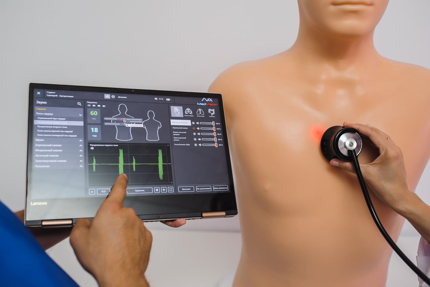
Furthermore, a digital ultrasound simulator provides highly objective, algorithmic assessment. Unlike a human instructor who might subjectively grade a student's scanning technique, the computing system inside the simulator meticulously tracks the precise angulation of the probe, the exact time taken to visualize the target organ, and the diagnostic accuracy of the student's final interpretation. This data-driven approach to training guarantees that every single graduate meets a strict, unyielding standard of clinical excellence.
Obstetrics and Transvaginal Ultrasound Simulation
The utilization of an ultrasound training simulator for medical students is absolutely paramount in the fields of obstetrics and gynecology. Performing a transvaginal ultrasound requires immense physical sensitivity, profound anatomical knowledge, and highly refined clinical skills. Utilizing live female patients for the early stages of learning in this highly intimate, invasive examination is often impractical, ethically fraught, and extremely uncomfortable for all parties involved. Therefore, implementing comprehensive ultrasound simulation models training utilizing a dedicated, highly realistic pelvic model ensures that medical students can repeatedly practice precise probe orientation and spatial image acquisition entirely risk-free.
A high-fidelity obstetric simulator allows instructors to program highly realistic fetal scenarios, including assessing fetal heart rate, measuring nuchal translucency, and identifying severe congenital anomalies. By rigorously engaging in ultrasound simulation models training, the learner develops the vital hand-eye coordination required to physically manipulate the transvaginal ultrasound probe while simultaneously interpreting complex diagnostic imagery on the monitor. This competency-based pedagogical approach ensures that when medical students finally enter the maternal-fetal medicine clinic, their foundational training on the simulator translates directly to compassionate, highly accurate, and professional patient care.
Cardiovascular Ultrasound and Echocardiography Training
Evaluating the dynamic, beating human heart in real-time is arguably the most cognitively demanding aspect of all sonographic education. To properly master transthoracic and transesophageal echocardiography, a premium ultrasound training simulator for medical students frequently incorporates a highly advanced, 3D holographic beating heart model. When medical students place the virtual transducer against the synthetic chest of the simulator, the simulation screen displays not only the standard 2D grayscale ultrasound view but also a simultaneous 3D anatomical cross-section.
This powerful dual-view simulation radically accelerates structural learning and spatial comprehension. Furthermore, just as the ultrasound simulator for emergency medicine prepares young doctors for blunt force trauma, a dedicated cardiac simulator prepares them for acute cardiovascular collapse. Students can relentlessly utilize the simulator to improve their ability to calculate ejection fraction, identify severe valvular regurgitation, and rapidly diagnose right ventricular strain. This highly repetitive, simulation-driven training guarantees that future practitioners of cardiovascular medicine possess the advanced diagnostic acumen necessary to manage life-threatening cardiac events with absolute confidence.
Global POCUS Integration and Standardized Testing
The global healthcare community is rapidly moving toward a future that requires mandatory Point-of-Care Ultrasound (POCUS) certification for almost all graduating physicians, regardless of their ultimate specialty. To achieve this monumental, standardized goal, the widespread integration of the ultrasound training simulator for medical students is the only logical logistical solution.
Professional regulatory boards and examination committees are increasingly relying on the advanced ultrasound simulator to conduct high-stakes credentialing and licensing exams. During a standardized test, every single candidate interacts with the exact same virtual phantom model, facing the exact same complex pathological scenarios. The simulator's internal computer provides an impartial, algorithmic grade reflecting the candidate's volumetric accuracy, tissue recognition, and total diagnostic time. This ensures absolute fairness and objectivity in medical credentialing, unequivocally proving that rigorous simulation is not merely a preparatory training tool, but the ultimate, undeniable arbiter of professional clinical competence.
Cognitive Load Theory in Ultrasound Education
The established psychological framework of Cognitive Load Theory heavily supports the mandatory, widespread adoption of the ultrasound training simulator for medical students. The real-world clinical environment of emergency medicine or the intensive care unit is inherently chaotic and overwhelmingly loud. If a novice is attempting to learn highly complex sonographic skills directly on a live, suffering patient, their working memory is completely overwhelmed by extraneous variables: the patient's palpable discomfort, the screaming alarms of the monitors, and the intense pressure of the supervising attending physician.
An advanced ultrasound simulator aggressively manages and titrates this severe cognitive burden. During early training, a clinical instructor can completely pause the simulation on the model, digitally highlight specific anatomical landmarks on the monitor, and allow medical students to process the complex geometric relationships without the crushing stress of a deteriorating human being. Once the foundational knowledge base is firmly established on the physical simulator, the instructor can progressively increase the difficulty of the scenarios, adding time limits and simulated patient distress. This stepwise, highly structured learning progression is exactly why the ultrasound training simulator for medical students is universally recognized as the single most effective educational methodology in modern healthcare.
The Economic Reality and ROI of Simulator Training
Acquiring state-of-the-art laboratory equipment for immersive ultrasound simulation models training requires a highly formidable financial investment from any academic teaching institution. High-end VR computing systems and ultra-durable, tissue-equivalent phantom models are undeniably expensive. However, a comprehensive, long-term financial analysis definitively reveals that the Return on Investment (ROI) for a modern ultrasound simulator is exceptionally robust.
Conclusion
In final summary, the massive, global integration of the ultrasound training simulator for medical students marks a definitive, irreversible, and highly positive evolution in modern medical education. The sheer technical and cognitive complexity of contemporary point-of-care sonography demands an uncompromising pedagogy that completely eradicates the inherent risks associated with early human experimentation.
By strategically implementing rigorous, standardized ultrasound simulation models training and widely deploying the high-fidelity ultrasound simulator for emergency medicine, academic universities establish an incredibly powerful, objective, and psychologically safe learning environment. This continuous, technology-enhanced practice ensures that every single graduate possesses the deeply refined spatial awareness and impeccable diagnostic mastery necessary to succeed. Ultimately, investing heavily in the modern ultrasound simulator guarantees that when these highly trained medical students finally face critical clinical emergencies in the real world, their sonographic response will be flawlessly swift, fiercely accurate, and profoundly life-saving.
References
- Ahmad, R., et al. (2020). "The role of high-fidelity ultrasound simulation in medical education: A systematic review." Journal of Ultrasound in Medicine, 39(12), 2285-2296.
- Gottlieb, M., & Bailitz, J. (2018). "Point-of-care ultrasound training in emergency medicine: The impact of simulation models." Annals of Emergency Medicine, 72(4), 450-457.
- Kaufman, J. (2019). "Cognitive load and simulation in healthcare: Optimizing the learning environment for medical students." Medical Education Online, 24(1), 162-171.
- Neff, L., et al. (2021). "Virtual reality and physical phantom models in obstetrics ultrasound training." Ultrasound in Obstetrics & Gynecology, 58(2), 211-219.
- Ziv, A., Rootenberg, L., & Macrae, H. (2016). "Simulation-based training in healthcare: A critical evaluation of the evidence base." Quality and Safety in Health Care, 15(suppl 1), i34-i43.
FAQ
What is an ultrasound training simulator for medical students?
An ultrasound training simulator for medical students is a highly advanced educational computer system or physical tissue phantom designed to flawlessly replicate the exact experience of performing a sonographic exam. It allows learners to safely practice scanning techniques, manipulate a virtual probe, and improve their image interpretation skills without ever practicing on live patients.
How does ultrasound simulation models training improve clinical skills?
Comprehensive ultrasound simulation models training dramatically improves clinical skills by providing infinite, risk-free repetition. Students use a physical model to build the necessary hand-eye coordination required to navigate complex anatomy. Because the simulator provides immediate, objective feedback, learners can rapidly correct their probe angulation and solidify their diagnostic knowledge base.
Why is an ultrasound simulator for emergency medicine critical for residents?
An ultrasound simulator for emergency medicine is absolutely critical because trauma and critical care require split-second diagnostic decisions. The simulator allows emergency medicine residents to repeatedly practice high-stress protocols, such as the FAST exam, in chaotic simulated scenarios. This rigorous training builds profound muscle memory, ensuring they can instantly identify internal bleeding during a real, life-threatening emergency.
Do these simulators really look and feel like real human tissue?
Yes. Modern physical ultrasound phantom models are explicitly engineered from proprietary synthetic polymers that perfectly mimic the acoustic impedance of real human flesh and bone. When a trainee presses an actual transducer against the model, the simulator yields to the pressure realistically, providing the exact tactile and visual feedback expected in authentic, high-level healthcare education.
Immerse yourself in a demo to see how MedVision transforms traditional learning into an engaging, interactive experience

Subscribe for the Latest News!